SINGEK BLOG: Tadpoles experiencing mass die-offs: Are protozoan parasites the cause?


As recently as 2006, travellers to Panama might have been treated to the spectacle of golden frogs brilliantly coloured against the deep green leafy background of the high mountains. Or perhaps they would have heard their calls rising up in a chorus with numbers reaching the thousands.
Today these frogs have disappeared from the wild and can only be found in small captive breeding colonies. The principle cause of their decline has been identified as the pathogenic chytrid fungus Batrachochytrium dendrobatidis (unwittingly spread by the same travellers that used to enjoy their spectacle).
Unfortunately, the plight of amphibians extends well beyond Panama. Nearly one-third of known species worldwide are threatened, while an additional 42 percent are reported as ‘declining in population’ (http://www.iucnredlist.org/initiatives/amphibians/analysis; accessed June 15, 2017). Like the Panamanian golden frog, many are imperilled by infectious disease.

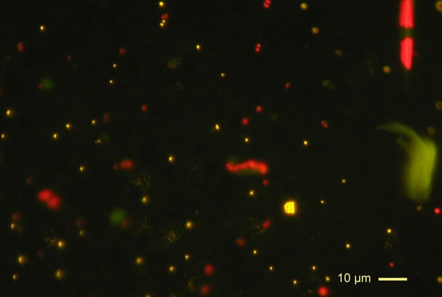
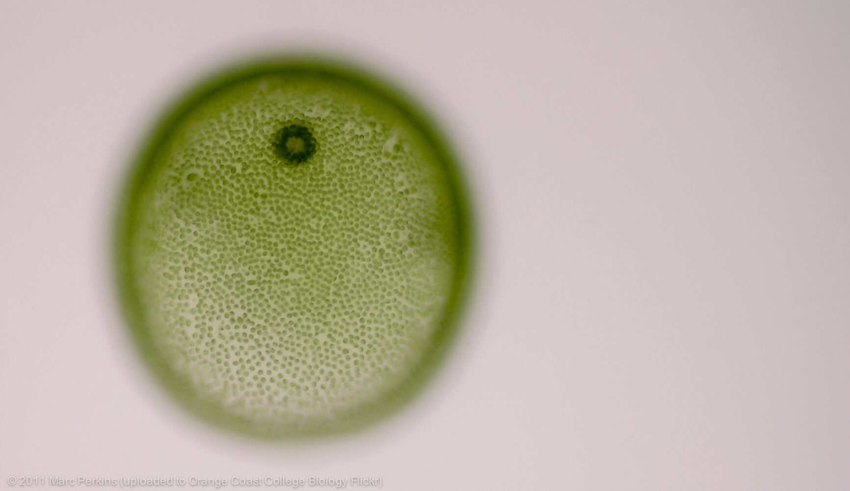
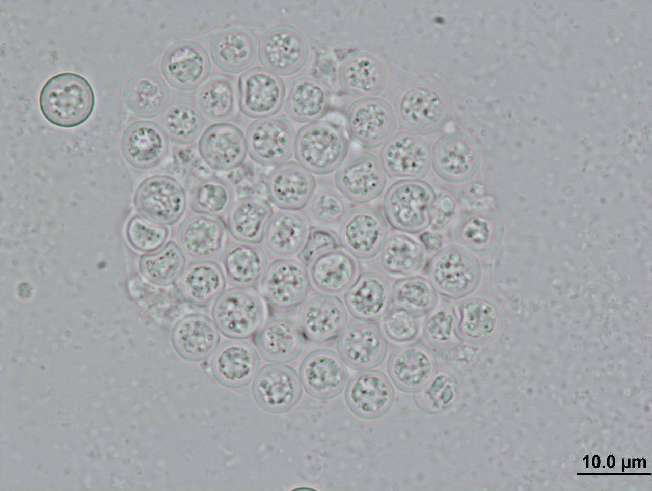
About the same time as the golden frog’s disappearance, researchers observed a mass die-off of southern leopard frog tadpoles from several ponds in Georgia, USA (Davis et al. 2007). They discovered that their body tissues, particularly their livers, had been infiltrated by a single-celled parasite similar to those in the genus Perkinsus (distant relatives of the agent of malaria) in both their appearance and genetic material.
Interestingly, another group of researchers later discovered Perkinsus-like genetic material in the tissues of tadpoles from 14 genera across a broad geographic range (Chambouvet et al. 2015); however, none of these tadpoles exhibited the symptoms that devastated the populations in Georgia. Indeed, it seems that the parasite can also cryptically infect amphibians without causing disease.
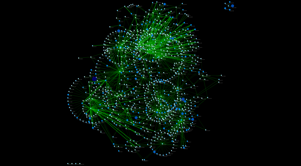
The outcome of infection is most likely determined by a complex interplay between the genes of the host and parasite, parasite levels in the environment, and extrinsic environmental factors. Over the next few years, my colleagues and I will begin to tease apart these dynamics.
To first establish disease causation, we will conduct infection experiments using different amphibian host species, inoculation methods and parasite burdens. These experiments will be followed by RNA analysis to identify the genes used by the parasite to facilitate infection. We also plan to conduct field studies of localities with reported die-offs to survey parasite levels and conditions that might be conducive to their pathogenesis.
Finally, we will use drug-sensitivity screening to identify compounds that inhibit growth of the parasite, providing a means to safeguard a captive breeding population should another species share the fate of the Panamanian golden frog.

About the author

Vanessa Smilansky – ESR 11

My MSc research was conducted at the University at Buffalo in Buffalo, NY, USA.  It involved the characterization of population genetic structure in a surface brooding Caribbean octocoral using microsatellite genotyping, genetic distance measurement and spatial autocorrelation. The research aimed to elucidate larval dispersal patterns, which we concluded occur on a fine-scale that varies temporally.

Following the completion of my degree in 2013, I worked as a genetics technician in the Washington Department of Fish and Wildlife in Olympia, WA, USA.  My work involved genotyping salmonids with microsatellite and SNP markers for use in genetic stock identification and parentage analysis.  Similar to the structure of the SINGEK network, the complexity of our projects and their far-reaching impacts led to a collaborative effort with other professional organisations both within and outside the agency.


SINGEK BLOG: Single and ready to mingle: The pros and cons of a multicellular lifestyle

SINGLE AND READY TO MINGLE: THE PROS AND CONS OF A MULTICELLULAR LIFESTYLE

Konstantina Mitsi

06/04/2018


Scanning electron micrograph of just-divided HeLa cells. Modified by Chiara Vitali.

Multicellularity is an exception, an extravagant and very expensive state, a luxury that very few organisms can afford from an evolutionary perspective. In addition, being unicellular allows you to be a cellular bachelor for life, independent and with no responsibilities towards anyone. Therefore, it still remains one of the greater mysteries of modern biology what convinced some cells to take the big step and to inseparably link their fates to others as part of the same entity.

Life started in the oceans and stayed there for a very long period of time. Imagine for a moment that you are a happy single cell swimming alone in the infinity of the oceanic depths and your only worries are to eat and reproduce. Your size matters; the bigger you are, the more options you have for your lunch menu and at the same time less possibility of being the lunch of somebody else. In a parallel universe, cells might just grow bigger and bigger without any limitations, but in ours the square-cube law doesn´t permit it. This principle states that, as a shape grows in size, its volume grows faster than its surface area. Consequently, a good solution for organisms wishing to grow larger was to stick together, specialize and cooperate.

Sharing the same living space with other cells can liberate you from the obligation to perform all the tasks by yourself; division of labour, known as differentiation, was a clear advantage of multicellularity that permitted organisms to develop elaborate structures. In addition, multicellular organisms can live longer and protect their propagules or gametes in a safe internal environment until they decide on the perfect time to reproduce – probably the most important advantage.

Of course, nature is not famous for being a charitable organisation, and the greater the benefits, the bigger the cost. Multicellularity implies close interactions and intercellular dependence; just a small amount of damage to a vital organ can lead to systemic failure, in other words it is a death sentence. Cells that join the multicellular brotherhood are also prepared to sacrifice themselves for the greater good at any time that is needed through a protocol known as programmed cell death or apoptosis; just take a look in the mirror and think that all your cavities were once filled with cells that voluntarily killed themselves to make some space. Last but undoubtedly not least, in some cases “rebel cells” arise and try to harm the community; this happens either because they are confused –as in autoimmune diseases- or because they become selfish – as in cancer.

To sum up, there is more than one way to do things and to be adaptive. Individuality offers independence whereas co-operation and specialisation bring flexibility. Whatever works!

About the author

Konstantina Mitsi – ESR 3

I received my BSc degree in Biology from National and Kapodistrian University of Athens in 2015. During my bachelor studies, I conducted a six month Erasmus Placement internship in the microbial biotechnology lab of Dr. Margarita Orejas in IATA-CSIC in Valencia (2012-20013), studying the rhamnose transporters of the filamentous fungus Aspergillus nidulans. In 2016, I acquired my MSc degree in Biodiversity (Evolutionary Biology) from University of Barcelona.


SINGEK BLOG: Software Development for Scientists

SOFTWARE DEVELOPMENT FOR SCIENTISTS

Imer Muhović

23/03/2018


Science is an inherently difficult field to work in. Today, more than ever scientists have to combine deep technical knowledge from different disciplines in order to create publishable work. Computational skills are expected from almost all scientists, however not everyone has acquired‚ them during their studies. This problem is amplified by the fact that a lot of scientific software is simply bad.

Whether this is by having poorly written documentation, buggy execution, complex installation procedures, no clear way of distributing software, or lacking any support from the authors, the developers of these tools are shooting themselves in the foot by limiting the use and impact of the tools that they have spent their valuable time on. The causes of such issues are up for debate, but what is certain is that all of us working in this field would benefit immensely from following some basic software development practices.

So the next time you are slaving away in front of your code editor, here are a few things to keep in mind:

  • You are not your end user.
    • The person who will be using your program will not have your level of expertise in software development and troubleshooting. They might be a Post-doc working in the same field, or they might be an undergraduate student from a distant country. Both of them should be able to use your tool with the same ease and understanding.
  • Has your tool ever been tested by anyone else?
    • We know it works on your machine, but that doesn’t mean much. For your tool to be acceptable for use in the real world you need to find a few testers to evaluate it. Make sure they are of different skill levels, ranging from completely non-technical to an expert in your field. Send them the installer and docs and ask them to actively try to break your tool. Leave them alone for a week, with absolutely no input from yourself. When you come back I guarantee they will have come up with cases that will break your tool that you would never have expected.
  • Does the documentation cover all scenarios the user could end up in?
    • Good documentation is invaluable and saves both you and the user from having to reply to each other’s emails. It must clearly convey how to install your tool, how to troubleshoot common issues, how to perform all of its intended actions, and would ideally offer a brief introduction to all the concepts you mention in it, with additional links to papers that offer more introduction to the field.
  • How will you distribute it?
    • You do not want to be the kind of author that distributes their code by email. GitHub accounts are free and allow you to keep your code in a visible, public place.
  • Will you actually support your tool after it is out?
    • You may have spent months cramped in front of a keyboard, painstakingly creating your masterpiece, but if you do not support your users, fix the bugs they discover, and offer improvements to your tool after it is out, you might just as well not have bothered working on it at all. In this case it will join the legion of orphaned GitHub repos, and papers with dead URLs, linking to promising tools, that never got a chance to shine because nobody was around to help it after it was published.

These are just common-sense tips, that any developer should follow, and I strongly believe that by making better tools we can lower the barrier of entry into science and give scientists more time to work on making new discoveries, and less time fighting against the tools they use.

About the author

Imer Muhović – ESR 15

I come from Sarajevo, Bosnia and Herzegovina. After obtaining my BSc degree in Genetics and Bioengineering at the International Burch University and halfway through my MSc, I started working as a QA Analyst at Authority Partners in Sarajevo. While outside my field of education, hard work and an analytical rigor obtained by a science education helped me excel in my work, and led me to spending two professionally satisfying years at that position.


SINGEK BLOG: Please, Mind The Gap

PLEASE, MIND THE GAP

Aurelie Labarre

09/03/2018


Photosynthetic picoplankton from the Pacific Ocean (off the Marquesas islands observed by epifluorescence microscopy (blue exciting light). Orange fluorescing dots correspond to Synechocococus cyanobacteria, red fluorescing dots to picoeukaryotes. Larger cells (e.g. diatom, upper right) can also be seen. Image original by Daniel Vaulot (CC BY-SA 2.5)

Reporting a lack of genomic diversity in the databases; a call to sequence more reference – and draft-quality genomes to significantly increase the understanding of marine microbial eukaryotes.

The oceans are Earth’s largest ecosystem, representing 71% of the planet’s surface. Despite its vastness, the diversity of the oceans’ life remains rather unknown. In fact, scientists estimate that less than 1% of the total species that it harbors are currently known. Among the lesser-known organisms in the ocean are the protists – single-celled eukaryotic organisms – denoting that they have only one cell, while the term ‘eukaryote’ means that each of these cells has a nucleus (and other organelles). Among the few well studied protists are the Radioloria (beautifully illustrated by Ernst Haeckel) and diatoms, which present a siliceous outer skeleton. But for long time they have often been overlooked and undersampled; they are now considered as a real neglected area of research and the reason are various. Above all: they are hard to study.

Classical taxonomy (the science which studies the classification of life) relies on morphological features of species. And, in spite of the fact that protists can be both plant-like or animal-like and share certain characteristics, they encompass an incredible diversity in their structure and function. However, many of the single cells are morphologically featureless; making it very difficult for a taxonomist task.

Another fundamental reason is that a large fraction of protists are unculturable, meaning they cannot be grown in a laboratory, hampering their study by scientists.

One such enigmatic uncultured lineage is named ‘MASTs’ (MArine STramenopiles). They represent a major community among aquatic protists. Considered as heterotrophs, they secrete enzymes that digest food externally after grazing on bacteria (phagocytosis). In fact, these tiny protists (up to 3µm) consume efficiently viruses, bacteria, picophytoplankton and are ingested mainly by other protists and small crustaceans. They are particularly adapted to this process as they possess special appendages, a long whip-like structure, used to create feeding currents that enhance prey capture. These heterotrophic protists are still hugely understudied and so cause a real taxonomic gap in our knowledge of the earth’s biodiversity.

In response, new technologies have facilitated genome investigation with the development of new techniques in environmental molecular sequencing. Single cell genomic analyses represent a broad and fast solution to build more reference databases for novel and uncultured species. In single-cell genomics, an individual microbial cell is isolated, lysed and the whole genome is amplified. By studying entire genomes rather than individual genes, single-cell contributions have a great potential to understand the full complexity of cells for novel microbes (suffering from a real lack of reference protist genomes) by revealing genome-wide properties to explore cell identity.

In this context, we aim to investigate ecological relationships and trophic role within the plankton community. However, the lack of systematic genome comparisons of marine microorganisms leads to a lack of understanding of marine biology in general. Hence, we would provide an effort in sequencing more heterotrophic flagellates with a focus on the MArine STramenopiles, which we believe will lead to a model for investigating links between diversity and ecological function in marine systems. Since the MASTs are divided into 18 sub-groups with the potential for associated ecological niches for each, recovering a large part of the MASTs distribution over their “family tree” will fill this gap in under-sampled areas and increase the taxonomic heterogeneity.

In pursuit of reconstructing total eukaryotic biodiversity, better efforts must be made to sample the whole of the ocean’s biodiversity by developing new techniques and methods. Among these are i) the power of collaborative open research and ii) increasing the general public’s interest, allowing them to perceive the crucial role of protists in our oceanic ecosystems.

About the author

Aurelie Labarre – ESR 1

I started my academic training in bioinformatics and genomics as a Bachelor and a graduate student at the University of Laval in Canada. A key part of my graduate work was studying the genomic evolution of eukaryotic micro-organisms within the Chlorophyceae lineage. To gain more direct work and research experience, I then moved to the UK to take part in a bioinformatics genome project of a Oomycete-related organism; followed by a metagenomic project in Norway in order to broaden my experimental and analytical skills. Through all these experiences, I have been fascinated by the world’s rich biodiversity and my progress in these research areas has inspired me to want to work more on the genomic diversity of marine protists.


SINGEK BLOG: No longer an imaginary world

NO LONGER AN IMAGINARY WORLD

Luis Galindo

22/02/2018


Single cell genomics give us a new view of microbial diversity

Mark Twain once said, “You can’t depend on our eyes when your imagination is out of focus”. The famous writer probably had other things on his mind when he coined this phrase, but I consider it as a good starting point for the request I am about to make.

My request is rather simple, I just want you to imagine. I want to imagine a world where your eyes just perceive a small part of a bigger world around you. Imagine that you are seeing through a small hole in your door. If I ask you now to describe what you are perceiving, what apparently is just a tree from your point of view, could actually be a whole forest, and what may look as one house, may actually be an entire town.

As a scientist, I have to constantly cope with this narrow perception of the world that I am trying to understand. I work with protists, small, unicellular eukaryotes. There is an astonishing diversity of those organisms, but we can obtain information of only a few of them. Even though culturing methods have been really helpful to get a general picture of their diversity, they only provide a narrow view. This image is even more coarse grained, since cultured microbes end up becoming monoclonal. This means, we end up with clones of a single individual cell, which clearly do not represent the diversity in nature. As a rule of thumb, only about 1% of all microorganisms are currently cultivable in a laboratory environment. Hence, the hole in my door is probably even smaller than the one you just imagined.

My line of work is key to get insights into this diversity of microorganisms, to clarify the relationships between all eukaryotes, to understand their origin, and therefore, our origin. Single-cell genomics is a recent tool that is giving us an opportunity to finally access this hidden diversity. In the past, we had to culture unicellular microorganisms in a lab before we could obtain information about their DNA. With single-cell techniques we are now able to get all the information we need from only one cell, allowing us to avoid the culturing step. This gives us a view of the microbial world closer to reality.

Single cell genomics allow us to take a drop of water from any pond around the globe, separate the microorganisms it contains and classify them individually according to their genetic information. With single cell genomics, we have a tool that gives us this information from each individual, with all their diversity included. This analysis method delivers an enormous amount of new data, and a new world of possibilities for us scientists, which just makes us rub our hands.

Nevertheless, single-cell genomics still need a lot of improvement. The method of amplification of DNA introduces certain biases to the analysis. The lysis of unicellular eukaryotic cells still needs improvement, and our ability to obtain and sequence genomes is clearly behind our ability to analyse them computationally. However, there is a new generation of researchers working every day to overcome these obstacles, researchers that keep on imagining. They imagine themselves not looking anymore through that little hole, they imagine themselves opening the door and finally getting a full picture of the diversity of the world which we are part of, a diversity that we are not even able to comprehend yet.

About the author

Luis Galindo – ESR 7

I am an Evolutionary Biologist original from the Canary Islands (Spain), and I have been selected by the SINGEK training network to work in the ESR7 project in Deep Eukaryotic Phylogenomics. I obtained my biology degree from the University of La Laguna in Tenerife. During my degree, and after I graduated, I worked with the Molecular Genetics and Biodiversity research group in the IUETSPC (University Institute of Tropical Diseases and Public Health of the Canary Islands).


SINGEK BLOG: The Broad Applications of Single Cell Technology

THE BROAD APPLICATIONS OF SINGLE CELL TECHNOLOGY

Atefeh Lafzi

08/02/2018


All multicellular life start from a single cell. As cells divide and differentiate , changes in transcriptional profile occur, leading to considerable phenotypical diversities forming different subpopulation of cells and tissues in living organisms.
Figure Credits: Tarryn Porter and Iain Macaulay, Sanger Institute.

Single cell technology – the technology that makes it possible to study individual cells rather studying cultured colonies consisting of thousands or millions of cells – has provided us a magnifier to look at cells one by one and to study them separately and of course more precisely!

So far under the SINGEK consortium, we have always been talking about single cells that are uncultured microbes or more specifically microbial eukaryotes. We have been talking about how these organisms are crucial for understanding evolution and the functioning of ecosystems. We also discussed the best methods to study these single cells, from procedures to collect the samples to the tools to define their position in a phylogenetic tree of life.

Single cell technology is being used beyond SINGEK in many different fields of biological research. Beside Microbiology and Evolution, research areas like Complex tissues, Neurobiology, Immunology and Cancer are among the top fields that single cell technology is being used. In the same ways that we are able to separate microeukaryotic single cell organisms, we are also able to separate and study tissue specific cells, neurons, sperm cells, oocytes and cancer cells. Nowadays many researchers are interested to take advantage of single cell technology to answer questions like:

  • How do tissues develop from stem cells? This can be done by tracking the path of individual cells while evolving to specific tissues like liver or muscle.
  • How heterogeneous is each tissue in our body which we maybe superficially consider as collection of identical cells?
  • How do embryos develop to give rise to a complete multicellular organism with different tissues, each specialized in a specific set of functions?
  • How do immune system specific cells react and behave in response to infections and other perturbations?
  • How do tumors evolve from individual, regular cells?

All these and many more questions are new approaches that single cell technology provides a reasonable amount of precision to study. The challenge right now is to improve this technology in terms of how to best separate cells, how to extract the DNA and RNA content from them, how to sequence their genetic information and how to analyze this data to answer new questions that this technology has made us able to think about.

As an Early Stage Researcher in the SINGEK project, I will be working on developing statistical and computational methodologies to address these questions. I will try to integrate machine learning algorithms as well as other statistical methods to better understand the data coming out of single cells, to increase the quality of the data and at the end to extract as much information as possible from this basic units of life.

About the author

Atefeh Lafzi – ESR 6

I am currently a PhD student in Biomedicine and work as SINGEK ESR 6 on development of methodology for single cell organism genome analysis in CRG-CNAG, Barcelona. I am interested in searching for procedures and data analysis methodology for single cell studies of species for which no reference genome sequences exist. Establishment of methodology for homogeneous DNA amplification from single cells, de novo genome assembly, RNA analysis and de novo transcriptome assembly are also among the areas I will work on during my PhD.


SINGEK BLOG: Filling the Gaps with dark matter – reconstructing the tree of life

FILLING THE GAPS WITH DARK MATTER – RECONSTRUCTING THE TREE OF LIFE

Max Emil Schön

05/01/2018


The tree of life as the scientist/artist Ernst Haeckel envisioned it in the late 19th century (bacteria and archaea were not known then as such); right: A current update of the tree of life using available genome sequences and covering a wide range of prokaryotes and several eukaryotic lineages.

The tree of Life as we currently display it, is full of gaps and missing parts, reflecting enormous evolutionary distances between organisms. These gaps make it hard to confidently make statements about their evolution. Luckily, we are living in exciting times, where environmental sequencing enables researchers to describe new orders and even phyla on a regular basis. It feels like a renaissance after the classical naturalists of the 18th and 19th century described many of the species known today. In the last year or two, several studies where published that exhibit an unprecedented magnitude of newly discovered organisms. Each of them presented hundreds or even thousands of complete genomes extracted from metagenomics sequences. Metagenomics means sequencing DNA from all the organisms present in a sample at the same time.

This so-called ‘microbial dark matter’ (because we did not know it existed before genome sequencing) can be used to fill the gaps in the tree of life. This can reduce the artifacts of phylogenetic inferences, which is the reconstruction of the evolutionary history between organisms from their genes and genomes. Our job is to make sure that the phylogenetic models we use integrate this huge number of newly discovered genomes. We are employing better models of sequence evolution but also integrate, for example models of protein family evolution or the evolution of protein structures. The results of this combined approach should be of greatest importance to the whole field of evolutionary biology. It will enable us to better understand microbial diversity and even shed light on parts of evolution that remain some of the greatest mysteries in biology, such as the origin of eukaryotes.
References

Haeckel, E. Generelle Morphologie der Organismen (1866); Hug, L. A. et al. A new view of the tree of life, Nature Microbiol. 1, 16048 (2016).

About the author

Max Emil Schön – ESR 5

After my undergraduate degree in geoecology, and especially after a Bachelor’s project on the phylogenetic relationships and diversity of fungi I choose to change my field of studies to bioinformatics and computational biology. I wanted to be able to thoroughly understand the tools I was using and develop improvements or new tools by myself. Nevertheless, as I was mainly interested in those methods as a way to understand relationships between organisms (from an evolutionary as well as from an ecological perspective), I did not want to abandon geoecology neither. At the University of Tübingen I’ve had the awesome possibility to study both degrees in parallel, and I learned a lot about bioinformatics as well as evolution and biodiversity during this time. The background from both my studies was applied during a joint Master’s project, where I evaluated bioinformatics methods for the assessment of species diversity in environmental DNA samples using metabarcoding and applied those methods to a next-generation-sequencing dataset generated from symbiotic fungal communities in roots and soil.


SINGEK BLOG: The Who with Whom? Marine Microbial Interactions Networks

THE WHO WITH WHOM?

MARINE MICROBIAL INTERACTIONS NETWORKS

Ina Maria Deutschmann

15/12/2017


This network contains dots (called “nodes”) and lines (called “edges”) combining two dots with each others. Each microbial species is represented by a node. If there is an interaction between two species, we connect the nodes by an edge. All nodes and edges combined constitute the marine microbial interaction network.

Just as we interact with other people, living beings, and nature, microbes also interact with their surroundings. To understand the marine microbial ecosystem, we need to understand these interactions. Phenomena like climate change can have a negative impact on this system. However, the marine microbial ecosystem is fundamentally important for example the food chain, oxygen production, and storage of carbon dioxide.

In my work, I use a mathematical construct that is helping us to order, structure, visualise, and ultimately understand interactions between microbes. We call this construct “network”: it contains dots (called “nodes”) and lines (called “edges”) combining two dots with each other. Each microbial species is represented by a node. And if and only if there is an interaction between two species, we connect the nodes by an edge. All nodes and edges combined constitute the marine microbial interaction network.

In order to construct such a network, we obtain water drops from the open ocean on a monthly basis. We check which microbes we find and measure how often they appear. This list is a time data set. There are programs that can use this set to predict which of the microbes are interacting with which other ones. One simple method is to compare trends. If there is an increase of one microbial species connected to an increase of another microbial species, there is probably an interaction. If there is a decrease of one microbial species connected to an increase of another microbial species, there is probably also an interaction. In both cases, the program will predict an interaction between the two microbes. We can draw an edge between the two dots representing both microbial species.

The problem with this approach is that these predicted interactions do not necessarily mean that there is also a real (true) interaction. In fact, the program predicts too many interactions of which we believe not all are true, e.g. we obtain networks which contain too many edges. These networks most likely fail to give an adequate overview of the ecosystem, and are not helpful to understand the ecosystem.

We can use environmental parameters to remove potentially false edges. Imagine two microbial species that grow in number when the temperature rises, and decrease in number if the temperature drops. Over the years, you could observe a pattern in which the number of those microbes rises in summer, and goes down in winter. Mathematically, the program would predict an interaction, but biologically that is not necessarily true, they maybe just both like warm temperatures.

Currently, we are working on a computer program that identifies such cases. In addition to the list of the number of microbes, we include information about the environment (e.g. temperature, salinity, or the amount of other substances such as chlorophyll and dissolved oxygen). For each edge, we determine how strong the interaction between the microbial species is, and we determine how much evidence we can find for the interaction being biologically not true by considering those environmental factors. If we believe that an edge is not representing a real biological interaction, it is removed from the network.

We have different possibilities to then proof that there is really an interaction between two microbial species. We can use “Single Cell Genomics” for example. In recent years, this field which investigates one single cell at a time has grown rapidly. Sometimes, researchers find that it is not possible to separate cells. Maybe because one microbe was eaten by a second microbe, or because they live in symbiosis in which they are maintaining a physical connection. These cases are strong indicators for true network edges with a real biological meaning.

There is also a lot of literature already published about specific microbial species and their interactions to other species. We try to automatically parse the literature and we are confident that a predicted network edge is true, if an interaction has already been described in those papers.

Marine microbial interaction networks can represent the Who with Whom in the ocean, and can help us to improve our understanding of these complex ecosystems. Our next step is to make predictions about what will happen if one specific microbial species suddenly disappears, e.g. dies out. How will this affect other microbes, the whole marine ecosystem, and ultimately also us humans? Will the death of one microbial species result in other microbes spreading? Or will this start a chain reaction leading to the death of microbes, and possibly fish or even mammals?

About the author

Ina Maria Deutschmann – ESR 2

I am interested in using mathematical approaches to answer biological questions. My interdisciplinary research combines molecular ecology, molecular evolution and computational biology with biomathematics.

Beginning 2015, I was a visiting academic at the Allan Wilson Centre, Biomathematics Research Centre (University of Canterbury in Christchurch, New Zealand). In 2016, I obtained the Master of Science degree in Biomathematics (Ernst-Moritz-Arndt University in Greifswald, Germany).


SINGEK BLOG: Cells as micromachines: there no better engineer than Nature

CELLS AS MICROMACHINES: THERE NO BETTER ENGINEER THAN NATURE

Jari Iannucci

20/10/2017


Volvox 100x magnified. Image original by Mark Perkins CC BY-NC 2.0

My career path is somewhat unusual. I studied engineering and now I do my PhD with microorganisms. I am amazed by the complexity and efficiency of living systems. No mechanical device and no super computer does even get near the intricate details and complex regulatory mechanisms microorganisms display

Microorganisms are all around us and even inside us by the billions. They cause devastating diseases and at the same time help in brewing beer and making cheese. I am particularly interested how microorganism respond to modifications in their external environment, or habitat. Usually, biologists would culture large amounts of microorganisms to study their behaviour in response to a certain perturbation. My approach is different. I am interested in understanding individual cells.

To study individual microorganisms, I am using a cross-over approach between biology and engineering. By using technologies used to create microchips, I am creating mini-habitats for single cells. One can imagine them as tiny aquariums, one million times smaller than the ones you would keep your ornamental fish in. They are called microfluidic chambers –or lab on a chip.

Just like you would be mesmerized by the colours, shapes and behaviour of the fish in your aquarium, I am fascinated by observing microorganisms under the microscope in their microfluidic chamber. How do they respond to perturbations, for example changes in nutrient supply or temperature? Does each cell show the same behaviour or are there individual differences?

Aquariums have been around for 2000 years and observing fish in them has never lost its fascination. Only now has it become possible to study individual microorganisms in their own little aquariums. Does this now make me an aquarist? Or am is still an engineer with an interest in microorganism?

About the author

Jari Iannucci – ESR 12

Last January I achieved a Master Degree in Engineering of Nanotechnology with honors at ’Sapienza University of Rome’, with an experimental dissertation on ’High slope biochemical gradient for cell migration studies’. This involved a one-year internship at Institute of Bioengineering of Catalunya (IBEC) in Barcelona.


SINGEK BLOG: A floating lab to understand our planet

A FLOATING LAB TO UNDERSTAND OUR PLANET

Laura Rubinat

24/11/2017


Someone took a photo of me standing in front of the Tara that night.

I remember spending a long afternoon at the faculty working with some colleagues. It was a day in October and the streets of Barcelona were quiet empty, probably it was getting late. As I left the university library, I said goodnight to everyone and made my way straight towards the Moll de la Fusta quay. Finally.

When I arrived at the harbour, I immediately spotted her. I could hardly take my eyes off her. That metallic hull. The wrapped up main sail and jib. The electric orange of her name on the bow. Tara. I’m not sure if there were other people around me or anything else going on, but I do remember feeling the magic in the atmosphere that night.

I got closer to the edge of the quay and I put my hand on top of her silvery skin: a skin that had swam the seven seas, had resisted the arctic cold and had carried a floating laboratory all around the planet to study marine ecosystems. Tara is a beautiful schooner of 400 square metres that feeds scientists with unprecedented data.

The most remarkable feature of our planet is that 70% of its surface is covered by water. Oceans, these vast extensions of water masses, move incessantly transporting nutrients and heat. They regulate climate and synchronise energy fluxes in perfect harmony. They make this pale blue dot a safe and habitable home for us. However, our understanding of the complex regulatory cycles of the oceans as well as of the life inhabiting them is still very limited. This is where Tara comes into play.

For more than 12 years, the foundation Tara Expeditions has been organising ocean sampling campaigns to push forward the limits of our knowledge on marine systems and climate change. Tara expeditions work more or less like this: a team of adventurous scientists sails towards places where they can extract water samples of biological interest, and they send the material they collect to the labs as quickly as they can. In these labs are scientists who will take care of storing samples in freezers and who will use them for any sort of analyses – like microscopic imaging or DNA extraction. And finally, at the end of the chain, there are dozens of research groups that will analyse the data obtained to try to answer biological questions.

In my case, I’m part of the ones who open the computer every morning and spend hours looking at this data to explore the ecological patterns of microbes. While I do that, sometimes my mind wanders back to that night at the quay. Back then, the Tara crew only spent a few nights in Barcelona after a long trip in the Mediterranean. Right now, the boat is embarked on a new adventure in the Pacific. There are no limits for this floating lab of 36 meters length. Equally, there is hardly a limit for our passion to make sense of the data coming out of those expeditions.

You can follow the Tara pacific mission here: http://oceans.taraexpeditions.org/en/parcours-tara/

Video to be embedded: https://www.youtube.com/watch?v=xJkUmn8MgM4

About the author

Laura Rubinat – ESR 8

In 2009, I started a BSc in Biology at the University of Girona. During my bachelor, I completed an internship at the European Molecular Biology Laboratory (EMBL Heidelberg, Germany), where I studied the evolution of microbial cooperation interactions. Thereafter, I joined the Molecular Microbial Ecology group at the University of Girona to carry out my bachelor’s final project, which consisted on establishing synthetic biofilms to investigate the effects of spatial organization on bacteria denitrification capacity. I soon became interested in the infinite possibilities that bioinformatic tools offer us to answer biological questions, and after graduating I enrolled in the Master’s programme in Bioinformatics for Health Sciences, jointly organized by the Pompeu Fabra University and the University of Barcelona. I completed my master’s thesis at the Institute of Marine Sciences (ICM-CSIC, Barcelona), focusing on a comparative study of picoeukaryotic and prokaryotic phytoplankton diversity in the global ocean.